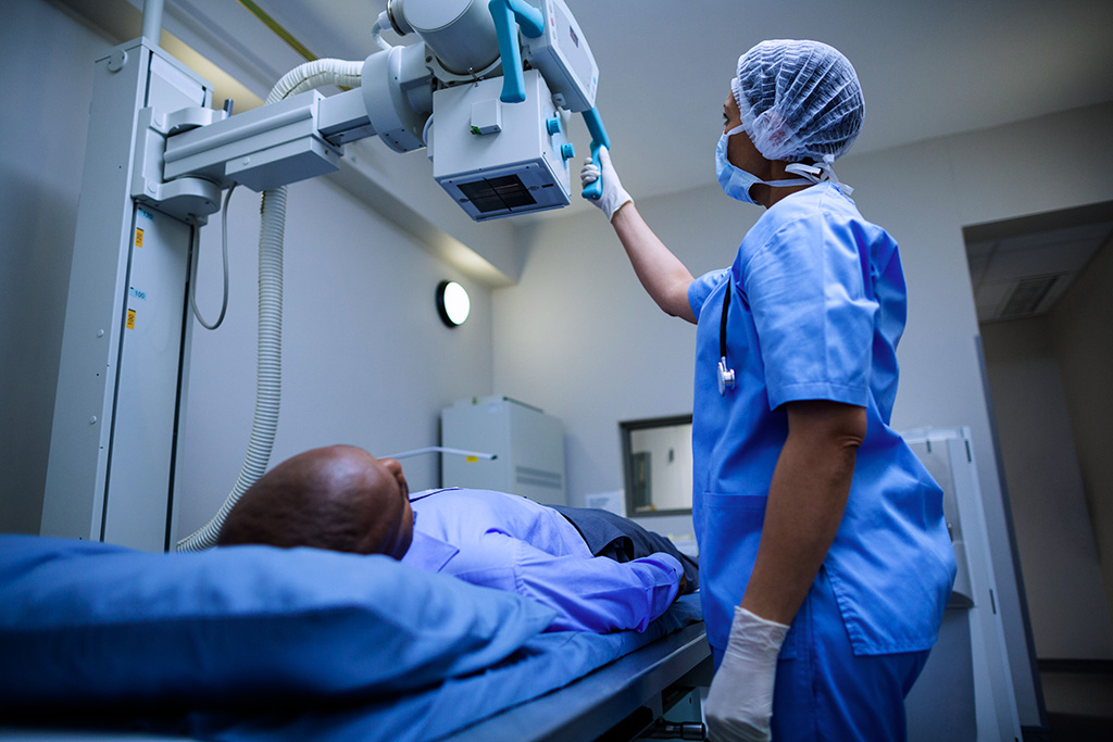
X-Ray
X-Rays are a common and quick diagnostic imaging method that uses low levels of radiation to capture images of the body’s internal structures. Dense materials, such as bones, appear white on the X-Ray, while softer tissues, like muscles, appear in shades of grey. X-Rays are often used to examine injuries, monitor medical conditions, or identify abnormalities. Depending on the procedure, you may be asked to stand or lie down for the scan. While generally painless, some positioning may cause mild discomfort. A medical referral is required for all X-Ray examinations. At BR Radiology Group, no appointment is necessary—simply visit your nearest location.
FAQs
No, appointments are not required for general X-Rays at BR Radiology Group. Our team strives to minimise waiting times, though there may be delays during peak hours.
Most X-Rays do not require any special preparation. You may be asked to wear a gown if necessary. To ensure an efficient process, consider wearing loose clothing without metal elements like zippers or buttons.
Please bring the following items:
- Your referral form from a healthcare provider.
- A valid Medicare or DVA card, if applicable.
- WorkCover claim details, if the X-Ray is for a work-related injury.
A qualified radiographer will guide you through the procedure, explain the process, and help you prepare. Depending on the area being scanned, you may lie on a table or stand upright. The scan itself takes only a few minutes. After the procedure, a radiologist will analyse the images and provide a report to your referring doctor.
BR Radiology Group bulk bills most Medicare-eligible X-Rays. However, some scans may involve additional costs. If applicable, our team will inform you of any fees before your appointment.
Reports are typically completed within 24 to 48 hours. For urgent cases, please inform the clinic staff so they can expedite the process in coordination with the radiologist.

